| Title | EFFECT OF PREVENTIVE TREATMENT WITH ALPHA-LIPOIC ACID AND VITAMIN E ON FREE RADICAL STATUS OF CHILDREN EXPOSED TO LOW-LEVEL IRRADIATION DUE TO CHERNOBYL ACCIDENT |
|---|---|
| Year | |
| Author | Ludmila G. Korkina and Igor B. Afanas’ev |
| Publisher | Russian Institute of Hematology for Children Leninskii pr |
EFFECT OF PREVENTIVE TREATMENT WITH ALPHA-LIPOIC ACID
AND VITAMIN E ON FREE RADICAL STATUS OF CHILDREN EXPOSED TO
LOW-LEVEL IRRADIATION DUE TO CHERNOBYL ACCIDENT
-Pilot Study-
Ludmila G. Korkina and Igor B. Afanas’ev
Russian Institute of Hematology for Children
Leninskii pr. 113 Moscow 117437, Russia
INTRODUCTION
The use of nontoxic antioxidants, free radical scavengers, and chelators in preventive medicine becomes one of the most promising methods in prophylaxis of “free radical pathologies”, i.e. the diseases associated with the overproduction or imbalance of free radicals in a human organism. At present, there are evidences of an important role of active oxygen species in pathogenesis of heart ischemic disease, arthritis, asthma, environment-associated diseases, etc.
Natural nontoxic vitamins possessing antioxidant and chelatory properties seem to be very perspective drugs for the prevention and treatment of free radical pathologies; therefore, their effects on damaging free radical processes are widely studied. Thus, it was shown that ascorbic acid and rutin (vitamin P) efficiently suppressed oxygen radical-mediated mutagenic effect of asbestos fibers on human leukocytes (1), lipoic acid and vitamin E inhibited lipid peroxidation, ischemia-reperfusion injury of organs and other free radical processes (2-4). The effects of vitamin E administration to patients with cancer, atherosclerosis, and Alzheimer’s disease have been also studied (5-8). Recently, we have shown the beneficial effects of long-term treatment with rutin of Fanconi anemia patients (9).
It is possible that the effect of irradiation on human organism is the most spectacular example of free radical-mediated injury as irradiation directly induces the formation of cascade of oxygen radicals (superoxide ion. O2-, hydroxyl radical, HO·, hydrated electron, eaq,etc.). These radicals interact with biological molecules resulting in the destruction of proteins, lipids, DNA, and various substrates and enzymes. Chernobyl accident and other accidents, which contaminated vast areas with radioactive materials, raise a new social problem of protection of million peoples living in contaminated regions with relatively low-level but permanent irradiation. Now, it becomes a world-wide problem, because low-level contamination is spreading unceasingly into new areas.
It is important that low-level irradiation may change the free radical status of a human organism enhancing oxygen radical production by cells and affecting endogenous antioxidant systems. Therefore, new methods of investigation of free radical status of an organism and of the protection against free radical damage induced by irradiation should be developed. We believe that one of the most important and perspective methods of protection of peoples living in contaminated regions are the long-term administration of nontoxic antioxidant vitamins.
In this pilot clinical trial we have studied the effects of the 28 days administration of two antioxidant vitamins, vitamin E and α-lipoic acid (Thioctacid®) as well as their combination on free radical status of children living in the area contaminated. With radioactive materials after Chernobyl accident. All children were living in regions with radioactive contamination equal to or more than 15 Ci/km2. They were practically healthy but characterized by the enhanced level of oxygen radical production in the blood. As α-lipoic acid possesses both antioxidant and chelatory properties, we hoped that the treatment of children with these two vitamins permitted to decrease the overproduction of oxygen radicals from the organism. The parameters of free radical status of these children before and after clinical trials were compared with those for children living in the non-contaminated area. It was found that the administration of lipoic acid and especially of the combination of lipoic acid and vitamin E normalized the free radical status of child’s organism.
MATERIALS AND METHODS
Children
Children were from the regions contaminated with radioactive materials after Chernobyl accident; they were living in the area with the density of radioactive contamination of 15-40 Ci/km2. After preliminary testing, 56 children with enhanced level of spontaneous luminol amplified CL of isolated leukocytes were selected for clinical trial and randomized into 4 groups:
Group A: 16 children were given 400 mg lipoic acid a day.
Group B: 14 children were given 400 mg lipoic acid + 200 mg vitamin E a day. Group C: 14 children were given 200 mg vitamin E a day.
Group D: 12 children (control group).
The age of the children ranged from 8 to 14 years (average age was 11, 4 +/- 2, 1 years). Selected children of both sexes (22 males and 34 females) were practically healthy without serious disorders in general biochemical and hematological parameters. For comparison, 23 healthy children of the same age range from the Moscow region were tested.
At the beginning and after the completion of clinical trial, pediatrician observation and all traditional hematological and biochemical analyses essential for the evaluation of living functions including a full blood count using the Coulter Counter technique (the hemoglobin content, erythrocyte, leukocyte, and platelet amounts and differential blood cell formula), the total protein, bilirubin, and creatinine contents and AST and ALT activities were performed for all the children participating in the trial. For measurement of the organism’s free radical status before and after the clinical trial, the intensities of spontaneous, PMA-, and latex-stimulated luminol-dependent CL by isolated leukocytes and the whole blood were measured. The corresponding analytical techniques are given below.
There were no drop-outs in the clinical trial due to illness or the toxic effects of drug administration. However, some children were excluded from the trial due to early departure from the holiday- house.
Blood Samples
Venous blood was drawn by disposed syringes early in the morning before breakfast into two tubes: the first was with 20 U/heparin and the second was without anticoagulant. 0,5 ml was collected into the first tube, and 9 ml was collected into the second one. Serum was obtained from the 9 ml blood sample by centrifugation and then was used for biochemical assay. The 0,5 ml blood sample was used for cell differential count.
Preparation of Leukocytes
The blood samples (1, 5 ml) were anticoagulated with 0,2 ml of heparin (20 U) in Hanks’ balanced salt solution (HBSS) and sedimented with 1,5 ml of dextran-metrizoate mixture (50 ml 6,2% dextrane/20 ml 38% metrizoate) at 25°C for 30 min. The cell-rich supernatant was centrifuged at 150 g for 10 min. Cell pellets were washed twice in HBSS. The final suspension of 2-3 x 10⁶ leukocytes was prepared in medium 199. The cells were counted with a microscope, and their viability was assessed by exclusion of 0,1% trypan blue dye. Cell differential count was confirmed by Giemsa staining.
Chemiluminescence Analysis
Luminol-dependent CL was measured on PChL-01 luminometer of USSR production. Leukocyte suspension (20 μl, 2 x 10⁶ cells/ml), luminol (50 μ M, final concentration), and 0,85 ml of HBSS were mixed in the 1 ml cuvette at 37°C. After 5 min, PMA (10ng/ml, Sigma) or the 0,1% suspension of latex (Sigma) in 0,9% NaCl solution was added, and the light emission was recorded continuously. The intensity of spontaneous CL and the difference between the maximal values of the cellular CL response to luminol and of spontaneous CL were measured.
Statistics
Medians, high and low quartiles, were calculated. Results were tested with the wilcoxon, Mann-Whitney U-test for significance for α = 0.05 and the box plot was used to demonstrate the results.
RESULTS AND DISCUSSION
The observation of children by pediatricians during and after the completion of the trial showed that there were no toxic side effects. Allergic reactions and other adverse events induced by the drug administration. All the children felt well during and at least 10 days after the trial. Biochemical and hematological analyses confirmed these observations. Pediatricians concluded that there was a very good tolerability to the drugs applied. More than that, there was significant beneficial effect of the administration of lipoic acid and the combination of lipoic acid and vitamin E on the liver and kidney functions.
A major danger for the children living in the regions contaminated with radioactive materials is the possibility of developing of pathologies which are initiated by free radicals. It is now well established that oxygen free radicals generated by irradiation in a human organism are able to damage DNA, proteins, lipids, and other biological compounds stimulating “free radical” pathologies, first of all, leukemia and tumors, depressing hematopoietic tissues, and inducing the disturbances of immune system. There are at least four major pathways of suppressing free radical processes and reducing the state of “oxidative stress”:
- Scavenging of free radicals by antioxidants and oxygen radical scavengers;
- The stimulation of the activities of antioxidant enzymes;
- Suppressing the activities of the enzymes catalyzing the production of oxygen radicals;
- Chelating and inactivating active ferrous ions, which catalyze the production of active hydroxyl and hydroxyl-like radicals.
It is known that lipoic/dihydrolipoc acid exhibits both antioxidant and chelating activities, while vitamin E (α-tocopherol) is a powerful natural lipid-soluble antioxidant. Therefore, we proposed that the administration of these nontoxic vitamins to the children exposed to permanent low-level irradiation could suppress damaging free radical processes in an organism by one or all the above pathways.
Free radical status of children before and after drug administration was estimated on the basis of the intensities of spontaneous, PMA-, and latex-stimulated luminol-amplified CL in leukocytes and whole blood.
Chemiluminescence Analysis
In this study we applied spontaneous, PMA-, and latex-stimulated luminol-amplified CL as an assay on oxygen radical production by isolated leukocytes. (A choice of the above stimuli was based on the fact that PMA activates the cells through a special receptor while latex is an agonist acting without any receptor). All four groups of children from the regions contaminated with radioisotopes exhibited before trial enhanced levels of oxygen radical production by leukocytes: the intensities of spontaneous, PMA-, and latex-stimulated CL were equal to the medians: 1014-1492, 13676-34151, and 27620-36843 counts/103 cells, respectively, against the medians: 902, 13521 and 35675 counts/103 cells for controls.
Thus, we found for the first time that the exposure of children to small doses of irradiation during 6 years results in a significant increase in oxygen radical production by blood phagocytes. (It should be noted that occasional measurements of luminol-amplified CL produced by the leukocytes of children from the same regions 2-3 years ago did not show such a big increase in oxygen radical production. Therefore, current CL measurements indicate a high risk of the development of free radical pathologies in the children living in contaminated regions more than 3-4 years).
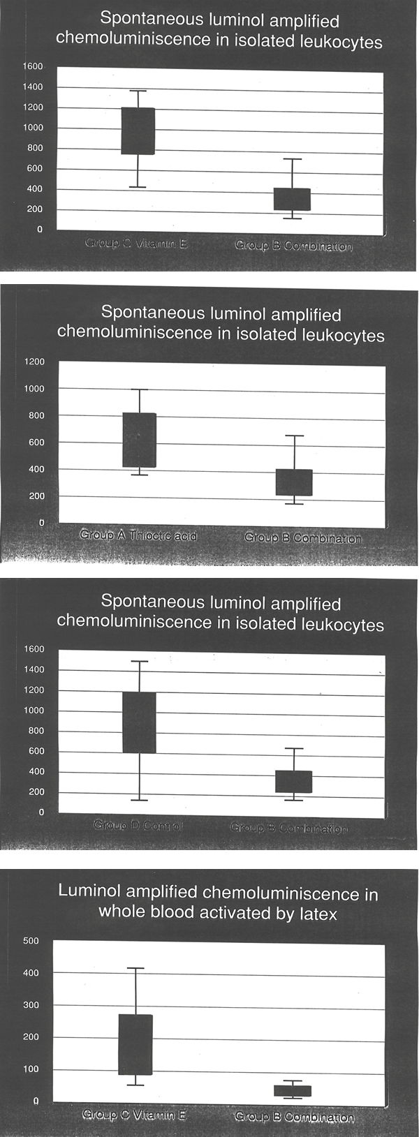
In the spontaneous luminol-amplified CL the combination of α-lipoic acid and vitamin E (Group B), was significant superior to vitamin E (Fig. 1) given alone (Group C) and to thioctic acid (Fig. 2) given alone (Group A) and to the control group D (Fig. 3). Thus, a maximal inhibitory effect on spontaneous oxygen radical production by isolated leukocytes was observed when the children were treated with the combination of 400 mg lipoic acid + 200 mg vitamin E. It is seen that after the administration of this combination or lipoic acid to children (Groups A and B) during 28 days, the level of spontaneous CL achieved a normal value.
It is important that the effects of separate administrations of lipoic acid and vitamin E (Groups A and C) were significantly smaller than that of their combination, vitamin E showing practically no effect on spontaneous CL. Thus, one may conclude that the combination of lipoic acid + vitamin E manifests a significant synergistic effect on spontaneous oxygen radical production by leukocytes in comparison with its components.
In contrast to spontaneous CL, lipoic acid and vitamin E given alone did not change significantly the levels of PMA-stimulated and latex-stimulated CL. We believe that the above findings can be explained as follows. Even the low but permanent doses of irradiation are apparently able to enhance spontaneous oxygen radical production by direct or indirect activation of dormant leukocytes. This phenomenon may be compared with weak inflammation. In accord with the above cited mechanisms, antioxidant vitamins may inhibit the enzymes catalyzed oxygen radical production. It is known that lipoic acid and vitamin E are not very active scavengers of superoxide ion. Therefore, we assume that their strong synergistic effect is mainly explained by the inhibition of activation mechanisms of leukocytes by irradiation.
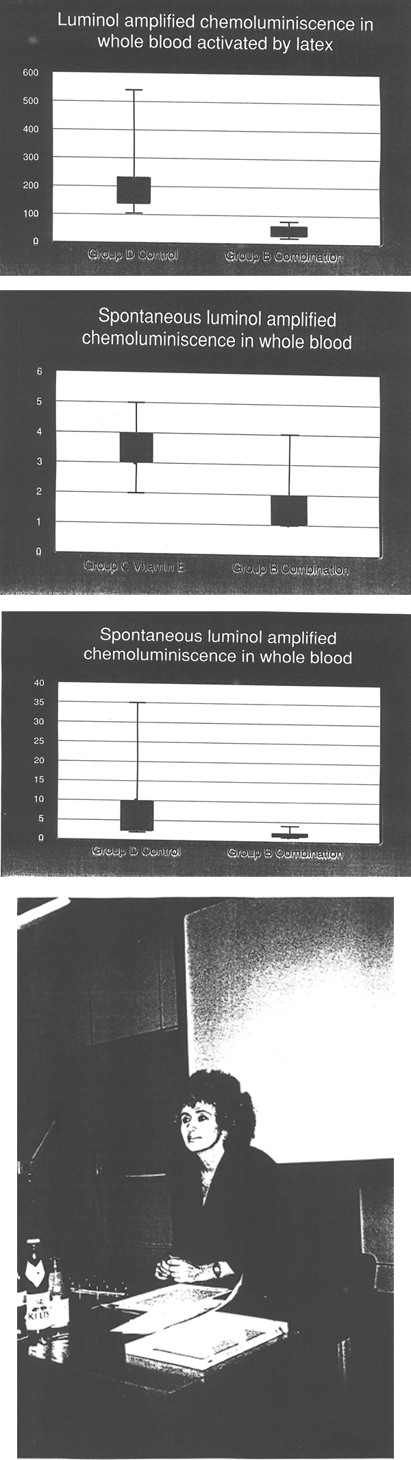
Both the soluble stimulus PMA and particular stimulus latex sharply enhanced oxygen radical production by leukocytes. It seems that lipoic acid and vitamin E were unable to interfere with the mechanism of PMA- and latex-activation. However, the last conclusion is only relevant to isolated leukocytes. In the spontaneous luminal-amplified CL without and with activation by latex in whole blood the combination of thioctic acid + vitamin E was significant superior to controls (Group D) and to vitamin E given alone (Group C) (Fig. 4-7). The study of whole blood showed that the combination of lipoic acid with vitamin E strongly inhibited both spontaneous and latex-stimulated luminal-amplified CL, the CL levels being reduced to normal values. We suggest that lipoic acid and vitamin E are able not only to inhibit the oxygen radical-producing NADPH oxidase of leukocytes but to enhance the oxygen radical- scavenging activity of serum.
CONCLUSIONS
- Administration of all drugs (vitamins), lipoic acid (400 mg a day), lipoic acid + vitamin E (400 mg and 200 mg a day), and vitamin E (200 mg a day) during clinical trial (28 days) resulted in no side adverse effects in all children.
- The administration of lipoic acid and the combination of lipoic acid + vitamin E resulted in a sharp decrease in oxygen radical production by isolated leukocytes and the whole blood.
- It is found for the first time that lipoic acid and vitamin E manifest a strong synergistic action on oxygen radical production by isolated leukocytes and in the whole blood. On these grounds, the combination of lipoic acid and vitamin E should be recommended for the treatment of children (and adults) with the enhanced level of oxidative stress, especially for the preventive treatment of children living in the area contaminated with radioactive materials and therefore exposed to permanent irradiation.
REFERENCES
- L.G. Korkina, A.D. Durnev, T.B. Suslova, Z.P. Cheremisina, N.O. Daugei-Dauge, and I.B. Afanas’ev, Oxygen radical-mediated mutagenic effect of asbestos on human lymphocytes: suppression by oxygen radical scavengers, MUTAT.RES. 265,245-253, 1992.
- K. Schmidt, Free radical diseases- prevention and therapy, in 2. INTERNATIONAL THIOCTIC ACID- WORKSHOP, ASTA Medical, Frankfurt am Main, 1992, pp. 15-23.
- L. Packer, New horizons in antioxidant research: Action of thioctic acid/dihydrolipoic acid couple in biological systems, IBID. pp. 35-36.
- H. Scholich, M.E. Murphy, and H. Sies, Antioxidant activity of dihydrolipoate and its dependence on α-tocopherol, BIOCHIM.BIOPHYS.ACTA 1001, 256-261, 1989.
- G.M. Paganelli, et al., Effect of vitamin A, C, and E supplementation on rectal cell proliferation in patients with colorectal adenimas, J.NATL, CANCER INST. 84, 47-51, 1992.
- B.Y. LeGardeur, S.A. Lopez, and W.D. Johnson, A case-control study of serum vitamins A, E, and C in lung cancer patients, NUTR.CANCER 14, 133-140, 1990.
- A.J. Verlangieri, Vitamin E and atherosclerosis, in VITAMIN E SIMPOSIUM, Henkel Co. 1990, pp. 8-10.
- Z. Zaman, S. Roche, P. Fielden, P.G. Frost, D.C. Niriella, and A.C.D. Cayley, Plasma concentrations of vitamins A and E and carotenoids in Alzheimer’s disease, AGE AND AGEING 21, 91-94, 1992.
- L.G. Korkina, E.V. Samochatova, A.A. Maschan, T.B. Suslova, Z.P. CHeremisina, and I.B. Afanas’ev, Release of active oxygen radicals by leukocytes of Fanconi anemia patients, J.LEUKOCYTE BIOL. 52, 357-362, 1992.
- E. Beutler, RED CELL METABOLISM, 1975, Grune & Stratton, N.Y., pp. 112-114, 115-117, and 69-71.
- CLIN.GUIDE TO LAB.TESTS, N.W. Tietz, Ed., Second Ed., 1990.
- A. Bast and G.R.M.M. Haenen, Regulation of lipid peroxidation of glutathione and lipoic acid: involvement of liver microsomal vitamin E free radical reductase, in ANTIOXIDANT IN THERAPY AND PREVENTIVE MEDICINE (I.Emerit, et al., Eds.) New York, Plenum Press, 1990, pp.111-116.
LEGENDS TO FIGURES
Fig. 1-3 Intensity of spontaneous luminol-amplified CL in suspension of isolated leukocytes after clinical trial. The median, low and high quartiles are given for each group. CL intensity is measured in counts/min 103 cells.
Fig. 4-5 Intensity of latex-stimulated CL in the whole blood. The median, low and high quartiles are given for each group. CL intensity is measured in mV/min 103 cells.
Fig. 6-7 Statistical treatment of data on spontaneous luminol-amplified CL in whole blood. The median, low and high quartiles are given for each group.
ACKNOWLEDGEMENTS
This work was supported by Henkel Co. We thank Dres. A. Koch and D. Meissner for helping in organisation of pilot trial and Professors A. Diplock, K. Schmidt, and F.J. Pellerin for fruitful and useful discussions.